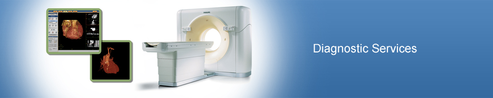
Diagnostic Services
Detection of Coronary Artery Disease (CAD)
 All of the organs and tissues in the body need a blood supply in order to function, as blood carries oxygen which is a source of energy. It is the heart's job to pump oxygen-rich blood through the huge network of arteries that extend throughout the body, including pumping blood into vessels that supply the heart muscle itself. These vessels, called coronary arteries, lie on the outside of the heart muscle.
- Coronary Artery Disease (CAD) or Ischemic Heart Disease (IHD) is a condition affecting these arteries that run on the surface of the heart and supply blood to the heart muscles. The most common cause of CAD is atherosclerosis, where fatty deposits including cholesterol and other fats, calcium and certain other elements carried in the blood build up as a plaque on the inside of artery walls. This tends to reduce the flow through the arteries and therefore, less oxygen and other nutrients reach the heart muscle. This can lead to problems ranging from pain in the chest to heart attack, heart failure, or rhythm abnormalities and sudden cardiac death. The chest pain (angina pectoris), or a heart attack (myocardial infarction) are manifestation of the same disease, the difference being that of severity. If it affects the arteries supplying the brain, a stroke (paralysis attack) may result.
-
 Angina usually occurs when the heart has to work harder than usual, as during exercise or emotion. There is pressure, tightness or pain in chest, arm, neck, back or jaw. The part of the heart muscle normally supplied with oxygen by the narrowed artery cannot get enough blood. With rest and medicines, the heart's demand for blood is met temporarily and so pain disappears. There is no permanent damage to the heart. With a heart attack, a narrowed artery is suddenly blocked completely by a clot forming at the point of narrowing. The part of the heart muscle supplied by that artery does not receive any oxygen and begins to die. This can damage the heart's ability to pump blood and causes chest pain, which does not disappear with rest. Symptoms of a heart attack may also include shortness of breath, sweating, weakness or dizziness.
Cardiac Imaging
X-Ray
- X-rays are a form of radiant energy, which penetrate various tissues of the body differentially, in the process capturing shadows and reflections on the photographic plate.
- The radiologist can view these on photographic film, on a TV or computer monitor. The films created by X rays show different features of the body in various shades of gray.
ECHOCARDIOGRAPHY DOPPLER
- Echocardiography is a specialized ultrasound imaging of the heart. It is done with help of a blunt flexible probe which is rotated over chest to send ultrasound waves to heart and receive the reflected waves to construct the image of heart and its functioning in real time. It is called a non-invasive procedure since it does not involve any injections.
- Patient will lie down
on theexamining table a technician places some lubricant and a transducer on the chest. The transducer beams high frequency sound waves at heart. This information is returned, or echoed, to the transducer and converted into a picture, which is recorded on videotape. Then the technician moves the transducer to several locations on chest until the picture is complete.
Cardiac MRI
- MRI is a method of producing extremely detailed pictures of body tissues & organs without the need for x-rays. MRI uses radiowaves & a strong magnetic field. In a strong magnetic field the radiowaves are directed at protons, the nuclei of hydrogen atoms. The protons are first "excited" & then "relaxed" emitting radio signals, which can be computer processed to form an image.
- MRI requires specialized equipment & expertise & allows evaluation of some body structures that may not be as visible with other imaging methods.
CT SCAN
- CT (Computed Tomography) is a diagnostic test that combines the use of x-rays with computer technology .A series of x-rays beams from different angles around the body are used to show cross sectional images of the patient's body. The images so obtained are assembled in a computer into a three- dimensional picture that can display organs, bones & tissues in great detail.
- In spiral CT, the examination table advances at a constant rate through the scanner gantry. While the x-ray tube rotates continuously around the patient, tracing a spiral path through the patient. This spiral path gathers continous data with no gaps between images.
- CT Angiography (CTA) is an examination that is used to visualize blood vessels in many areas of the body including the brain, kidneys, pelvis and the arteries serving the lungs. Compared to catheter angiography, which involves injecting contrast medium into an artery CTA is much less invasive & a more patient friendly procedure; contrast medium is injected into a vein rather than an artery.
Heart Rhythm Disorder
ECG
- An electrocardiogram (ECG or EKG) is a recording of the electrical activity of the heart.
- Each heartbeat is caused by an electrical impulse that causes the heart to squeeze. The ECG records the electrical pattern of the heartbeat.
- Many different diseases and conditions affect the ECG pattern. Small, sticky electrode patches are placed on chest, wrists, and ankles.
- These patches are connected to a machine that records the electrical activity of the heart.
- The recording is printed on paper for doctor to interpret.
HOLTER MONITORING
- This is a small monitoring device, which continuously records the ECG for 24 hours while patient continues his daily routine activities without remaining confined to bed.
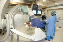
Electrophysiology Study
- An electrophysiology (EP) study finds the source of abnormal heart rhythms.
- Cardiac catheters and computers generate electrical impulses, and the speed at which the impulses travel within the heart is measured. These measurements help to locate impulses that are in the wrong place, as well as identify abnormal rhythms.
- The test takes about an hour and is not painful.
- It is a diagnostic procedure to identify the cause for irregularity in the rhythm of the beating heart.
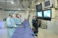
Ablation
- It is a curative procedure that corrects the arrhythmia identified. It involves delivering current at a particular radio frequency to break the abnormal circuit in the heart, returning it to its regular rhythm. BHIRC is one of the few hospitals in India to offers this therapy.
Cardiac Pacemaker - It is also a curative procedure performed on patients with abnormally slow heartbeat. It involves inserting wires into the heart chambers and regulating the heartbeat by delivering current from an external pacemaker placed under the skin.
Automatic Implantable Cardio-verter Defibrillator (AICD) - It involves implanting the AICD beneath the skin to monitor the rhythm of the heart and to deliver a carefully timed electric shock automatically, for life threatening rhythm disturbances.
Lung Function Test
PULMONARY FUNCTION TEST
- PFT are a group of tests that are performed to assess the functions of the lungs. Measuring the volumes during different phases of breathing, adequacy of airflow in and out of lungs and expanding capabilities or elasticity of lungs can assess the
lung functions.
- Tests can be performed not only to assess the breathing volumes and flows, but also the adequacy of oxygenation of blood that lungs are supposed to carry out.
- The major types of pulmonary function tests include spirometry, measurement of lung volumes, and quantization of diffusing capacity using small quantity of carbon monoxide.
- Measurements of maximal respiratory pressures and forced inspiratory flow rates are also useful in specific clinical circumstances Evaluation of lung functions is important in many clinical situations, both when the patient has a history or symptoms suggestive of lung disease, and when risk factors for lung disease are present, such as cigarette smoking.